About CXRML Classifier
Welcome to CXRML Classifier (Chest X-ray Machine Learning Classifier), a prototype solution aiming to assist medical professionals in diagnosing various pulmonary conditions through advanced image analysis. This application leverages machine learning algorithms to provide accurate and timely insights from chest X-ray images. Below is an overview of the application, its capabilities, and the technology that powers it.
Summary
CXRML Classifier is a diagnostic assistant for healthcare providers, utilizing DenseNet-121, a Convolutional Neural Network (CNN), to analyze chest X-rays. The application offers high accuracy, real-time analysis, and an intuitive user interface. CXRML Classifier includes explainable AI features, allowing users to understand the model’s decision-making process by highlighting significant regions in the X-rays. CXRML Classifier’s mission is to provide healthcare providers with diagnostic tools that improve patient outcomes. This application aims to bridge the gap between traditional radiological practices and modern machine learning techniques.
How It Works
This application is built on a sophisticated machine-learning model trained on thousands of chest X-ray images labeled as “Covid”, “Normal”, or “Pneumonia”. The model is designed to identify patterns and anomalies in these images, helping to detect the above-mentioned conditions. The X-ray dataset used to train the model was a compilation of roughly 6,000 Chest X-ray's on Kaggle from various sources of patients diagnosed with either Covid-19, Pneumonia, or normal lung health. The first step of this project was to split this data into training, validation, and testing datasets of which a 70%, 20%, and 10% portion of the data was used for each dataset respectively. Here is a detailed breakdown of how the application operates:
- Image Upload and Preprocessing: Users can upload chest X-ray images directly through the user-friendly interface. Once uploaded, the image undergoes preprocessing, where it is resized, normalized, and prepared for analysis. This step ensures that the input data is standardized, allowing the model to perform optimally.
- Feature Extraction: The core of the application lies in its ability to extract meaningful features from the X-ray images. Using DenseNet-121, a Convolutional Neural Network (CNN), the model scans the image to identify key patterns, textures, and shapes that may correlate to the presence of disease. These features are then fed into the model’s decision-making process.
- Prediction and Analysis: After extracting features, the model evaluates the image against its trained dataset to make predictions. The application identifies whether the X-ray shows signs of conditions like Covid-19, Pneumonia, or normal lung health. It also highlights the areas of the image that contributed most to the prediction, providing a visual explanation for the diagnosis.
- Results and Interpretation: The application presents the results in an easy-to-understand visual format. Users receive a clear prediction, such as "Covid", "Pneumonia", or "Normal". Alongside this prediction, the application highlights specific regions of the chest X-ray based on a threshold mechanism, which can be adjusted by the user. This highlighting is achieved through a technique called Guided Backpropagation. Guided Backpropagation works by calculating the gradients of the predicted class score with respect to each pixel in the input image. These gradients are obtained through a process called backpropagation, where the model computes how changes in each pixel would affect the final prediction. The gradients indicate how much each pixel in the input image contributes to the model’s decision. By using these gradients, the application can identify and highlight the regions in the X-ray image that are most significant for the prediction. When the user adjusts the threshold slider, the application highlights only those pixels where the gradient values exceed a certain percentage (controlled by the user) of the maximum gradient. This means that only the most significant pixels (those that contributed most strongly to the prediction) are highlighted. A lower threshold will result in more pixels being highlighted, showing a broader area of the image that includes less critical regions, while a higher threshold will narrow the focus to only the most relevant areas. This interactive feature provides users with the ability to explore different levels of model confidence, offering deeper insights into which parts of the X-ray were most influential in determining the diagnosis. The highlighted pixels serve as a visual representation of the areas that the model considered most indicative of the condition being predicted, helping to enhance the interpretability of the AI model’s decision-making process.
Key Features
CXRML Classifier offers several key features that if refined, could make it a useful tool for radiologists and healthcare providers:
- High Accuracy and Reliability: Trained on a diverse and comprehensive dataset, the model achieved a balanced accuracy of over 90% when classifying Chest X-rays of people with Covid-19, Pneumonia, or normal lung health.
- Real-time Analysis: The application provides results in real time, enabling quick decision-making in critical care settings.
- User-friendly Interface: The application was designed with ease of use in mind. The interface is intuitive, allowing users to upload images, view results, and interpret findings without needing extensive technical knowledge.
- Explainable AI: The application incorporates explainable AI techniques, providing users with a visual explanation of the model’s decision-making process. This transparency helps build trust in the AI’s predictions that could support clinicians in their decision-making.
The Technology Behind The Application
This application is powered by advanced machine learning technologies and frameworks, aiming to provide high performance and accuracy:
- Important Frameworks and Libraries: The training of the machine learning model for this project was conducted using the PyTorch framework. To optimize the model, Optuna was used for hyperparameter tuning, fine-tuning parameters such as learning rate and weight decay during the training process. This optimization aimed to achieve an optimal bias-variance tradeoff in the model. Additionally, scikit-learn's StratifiedKFold method was used for cross-validation, helping to mitigate overfitting while ensuring that each fold maintained the same distribution of class labels, thereby providing a balanced and thorough evaluation of the model's performance. Scikit-learn was also used to calculate the balanced accuracy
- Convolutional Neural Networks (CNNs): At the heart of the application is a CNN, a type of deep-learning model specifically designed for image processing. CNNs excel at recognizing patterns in visual data, making them ideal for analyzing chest X-rays.
- Transfer Learning: Transfer learning techniques were employed to enhance the model’s performance. By leveraging pre-trained models and customizing the final layer of DenseNet-121, the model was able to achieve a balanced accuracy of over 90% even with a relatively small dataset.
- Data Augmentation: To improve the robustness of the model, data augmentation techniques were used during training. This involved creating variations of the training images (e.g., rotations, flips) to expose the model to a wider range of scenarios.
- Guided Backpropagation: To provide visual explanations for the model’s predictions, Guided Backpropagation, a technique that finds the regions of the X-ray that were most influential in the decision-making process, was used.
Application Architecture
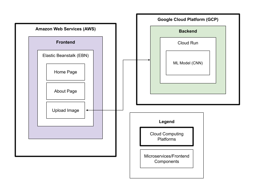
If you want to read more about this application's architecture, refer to the architecture overview here: CXRML Classifier Architecture Overview
Future Improvements
In future versions of this application, a large improvement that could be made is the usage of lung segmentation. Segmenting the lung out and having the Machine Learning model focus on that area would increase accuracy and relevance when detecting pulmonary conditions. This addition would also increase the reliability of the interactive gradient display.
Contact Me
Any inquiries and feedback from the medical community, researchers, and potential collaborators are welcome. If you have any questions, suggestions, or would like to learn more about this application, please contact me at justin.sy.huang@gmail.com.
To view the complete code for this project, refer to: GitHub Repository Link
My LinkedIn: linkedin.com/in/JustinHuang05
My GitHub: github.com/JustinHuang05